The Future of AI and Neuroimaging: A View from Our Founder Dr. Mark Haacke
By: Karen Holzberger, President & CEO of SpinTech MRI
Medical technology is advancing faster than ever before, including the world of MRI. MRI technology as we know it is less than 50 years old, but the implications of recent advances are astounding. What if we could complete a full standard clinical brain MRI in less than 10 minutes? What if we could eliminate the need for additional specialized patient scans? What if we never had to worry about a lack of data acquired through current scans?
The day is coming when a single neuroimaging protocol can create perhaps 20+ contrasts, half of them quantitative, in every scanning session. Rather than choosing which protocol to run, radiologists will simply choose which processed images to review, as all the relevant data has already been collected. Meanwhile, AI programs in the background will aid in analyzing all the collected and processed data, searching for biomarker abnormalities that warn of current clinical problems or future risks.
The Importance of MRI Data Standardization
The standardization of these new comprehensive protocols allow radiologists to compare data from different sites or scanners, regardless of manufacturer, to map out changes in the patient’s condition. SpinTech’s STAGE (STrategically Acquired Gradient Echo) software gives technicians a glimpse into the future of MRI. The images below represent one individual scanned in multiple cities on different systems and with several manufacturers. High-resolution scans can be achieved in just 8-10 minutes, and a slightly lower resolution in just 4-5 minutes.
-

Figure A: Dr. Wu Bo scanned on 7 different 3T magnets (5 Siemens, 2 Philips).The consistency is evident across scanners and across sites. -

Figure B: Average of the images over all 7 sites after co-registration. The high data quality provides sharp, clear images even after co-registration.
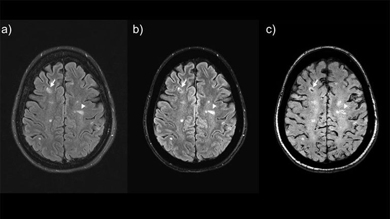
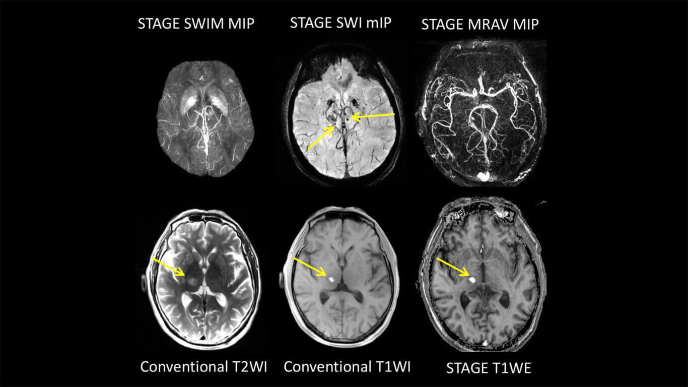
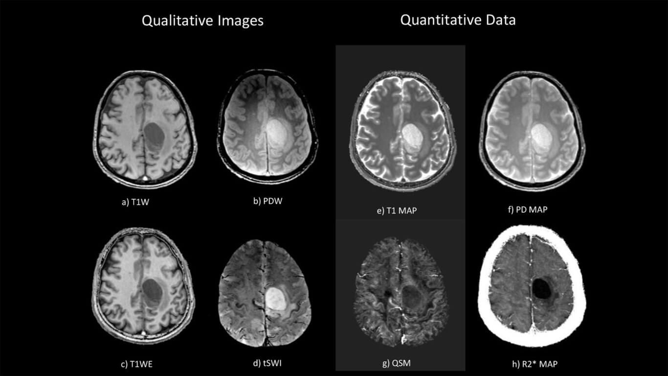
These new protocols provide T1, spin density and susceptibility information through a variety of contrasts as well as the ability to segment and quantify gray matter, white matter and cerebrospinal fluid (CSF). This data can be used to produce high contrast T1W images, susceptibility weighted images, T2* maps, susceptibility maps, and many other types of contrast including simulated FLAIR and simulated T2W imaging in less than 10 minutes.
Our Solution: STAGE MRI Software Innovation
With the advent of compressed sensing techniques, current imaging with STAGE may be able to produce good results in half to three-quarters the current times. We imagine that in the near future, if DTI, fMRI, MRS or a high-resolution T1 map are needed, they will require approximately 5 minutes each. This means a total time of 30 minutes to complete all necessary neuroimaging protocols.
The STAGE protocol currently produces 15 to 20 different types of images, depending on your choice of contrasts. Each data set is 64 slices with a matrix size of 512 x 512 (maximum) with both magnitude and phase data, totaling 0.64 Gigabytes. If you add in all orientations for arbitrary viewing in 3D, it amounts to roughly 2 Gigabytes of data per scan.
Most radiologists don’t have time to view and process such a significant amount of data, nor is an algorithmic approach available yet to analyze all the data quantitatively. Still, combining this data with clinical information using AI will open the door to finding image features that correlate with diseases, improving diagnosis on current conditions while detecting markers denoting future outcomes.
To effectively utilize this AI methodology, one needs a standardized imaging protocol to be successful. This is yet another way that STAGE plays a key role in the future implementation and validation of AI outputs.
Addressing Real-World Clinical Challenges
The importance of rapid, quantitative, and standardized multi-contrast imaging moves beyond the world of research and into concrete clinical applications. The implications of this technology solve critical clinical challenges faced by radiologists and neurologists.
Each year, over 10 million patients in the U.S. suffer from neurological conditions such as stroke, traumatic brain injury, dementia, Parkinson’s Disease, Multiple Sclerosis and brain tumors. This number only continues to rise, driven by an increasing occurrence of disease and an aging population. Quickly and accurately quantifying neurological biomarkers is critical to proper diagnosis and treatment.
For example, detecting micro-bleeds in TBI or stroke may impact whether or not a patient receives proper, timely treatment. Iron concentration in deep brain structures is shown to be a key biomarker in diagnosing and treating Parkinson’s. However, current imaging techniques are not capable of identifying and quantifying these biomarkers in a clinically meaningful timeframe and within existing workflows.
Moreover, radiologists already face strained workflows and have limited time to dedicate to each case. They’re under pressure to increase throughput and revenue while improving diagnosis accuracy and quality decision-making, and with conventional imaging techniques, these challenges are nearly impossible to overcome.
That’s why we created STAGE. We’ve created a solution that allows radiologists to increase clinical throughput to drive additional revenue, improve diagnosis while reducing read-times, and improve workflow through enhanced, comprehensive data. Additionally, having access to advanced, diagnostics can improve referral rates by advertising a higher standard of care. It also reduces the need to rescan patients, improving patient satisfaction and overall decreasing operating costs.
Applications of STAGE Imaging
Through our work with over 50 global research partners, we’re continually developing and expanding clinical use cases of STAGE imaging and some of its early AI applications. Here are a few of those early tools.
*The following tools have not been evaluated by the FDA and are currently for research purposes only.
Cerebral Microbleed Detection

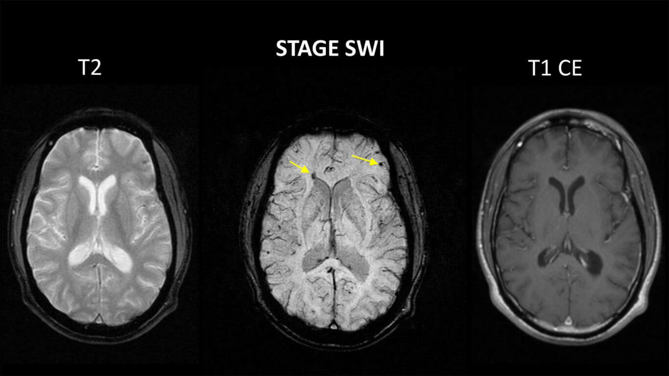
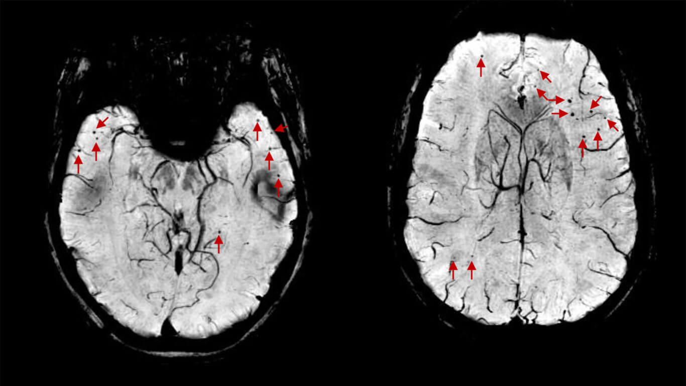
Early and accurate detection of cerebral microbleeds (CMBs) is critical for proper diagnosis and treatment of a range of conditions including stroke, TBI and dementia. For example, knowing the number, size, and location of CMBs in TBI is directly correlated with cognitive outcomes and treatment decisions. CMBs are also an important marker in stroke, showing signs of hemorrhagic stroke and influencing the administration of anti-platelet therapies. In dementia, CMBs are an important marker for predicting cognitive decline and assessing disease progression. However, CMBs can be time-consuming to identify manually and are often missed or misidentified as calcifications or blood vessels.
Using STAGE data we can apply machine learning and deep learning tools to automatically detect and quantify bleeds, showing the number, size, and location. Through comprehensive quantitative reports that are sent back to the PACS automatically, clinicians can now quickly and confidently assess a patient’s condition and make better treatment decisions as a result.
To learn more about this tool, and its clinical sensitivity and specificity, read our recent paper in Neuroimage: https://www.sciencedirect.com/science/article/abs/pii/S1053811919304380
Neuromelanin Quantification in Parkinson’s Disease

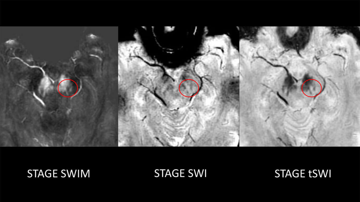
Parkinson’s Disease is one of the fastest-growing neurological disorders with over 6.2 million patients worldwide in 2015. Based on current trends, that number is expected to double by 2040. There are currently no definitive laboratory tests for Parkinson’s, but advances in MRI techniques have opened up new horizons in establishing a clinical imaging biomarker. Specifically, the concentration of neuromelanin in the substantia nigra and of norepinephrine in the locus coeruleus have been shown to correlate with Parkinson’s Disease. Visualizing and quantifying the Nigrosome-1 in the substantia nigra also provides important clinical information for diagnosis and monitoring.
Using STAGE data, we can accurately and automatically quantify these important biomarkers. Through our work in developing the Parkinson’s Disease Imaging Consortium in China, we have already scanned more than 500 PD cases and 500 healthy controls, leading to an age-adjusted normative database. This allows us to evaluate a patient’s condition and disease progression relative to known populations, while also differentiating Parkinson’s Disease from other movement disorders.
Our goal is to image an additional 500 cases each for Parkinson’s patients and healthy controls in the next year, first in China and now starting in the United States. These scans will further strengthen this tool by helping to establish reliable clinical biomarkers for this challenging disease.
To learn more, we invite you to read our most recent paper on this tool, which was featured on the cover of the September 2020 issue of Neuroimage: https://www.sciencedirect.com/science/article/pii/S1053811920304213
Learn More: Clinical Use Cases of STAGE Imaging
Beyond these early AI applications, we have also established clinical use cases for STAGE across a range of conditions. To learn more about these individual applications, please visit the links below:
Getting Started With STAGE
Whether you’re interested in supporting your existing research, exploring new frontiers, or simply improving your standard clinical imaging, we invite you to join us as we push this important technology forward. Reach out to schedule a demo and see what the data looks like on your own systems.
