STrategically Acquired Gradient Echo (STAGE) imaging, part I: Creating enhanced T1 contrast and standardized susceptibility weighted imaging and quantitative susceptibility mapping.
By: Karen Holzberger, President & CEO of SpinTech MRI
Abstract
Purpose:
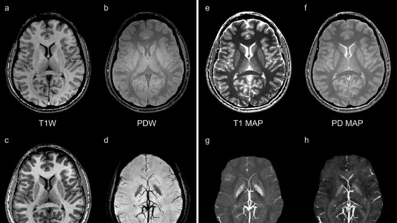
To provide whole brain grey matter (GM) to white matter (WM) contrast enhanced T1W (T1WE) images, multi-echo quantitative susceptibility mapping (QSM), proton density (PD) weighted images, T1 maps, PD maps, susceptibility weighted imaging (SWI), and R2* maps with minimal misregistration in scanning times less than 5 minutes.
Methods:
Strategically acquired gradient echo (STAGE) imaging includes two fully flow compensated double echo gradient echo acquisitions with a resolution of 0.67×1.33×2.0 mm3 acquired in 5 minutes for 64 slices. Ten subjects were recruited and scanned at 3 Tesla. The optimum pair of flip angles (6o and 24o with TR = 25 ms at 3T) were used for both T1 mapping with radio frequency (RF) transmit field correction and creating enhanced GM/WM contrast (the T1WE). The proposed T1WE image was created from a combination of the proton density weighted (6o , PDW) and T1W (24o ) images and corrected for RF transmit field variations. Prior to the QSM calculation, a multi-echo phase unwrapping strategy was implemented using the unwrapped short echo to unwrap the longer echo to speed up computation. R2* maps were used to mask deep grey matter and veins during the iterative QSM calculation. A weighted-average sum of susceptibility maps was generated to increase the signal-to-noise ratio (SNR) and the contrast-to-noise ratio (CNR).
Results:
The proposed T1WE image has a significantly improved CNR both for WM to deep GM and WM to cortical GM compared to the acquired T1W image (the first echo of 24o scan) and the T1MPRAGE image. The weighted-average susceptibility maps have 80 ± 26%, 55 ± 22%, 108 ± 33% SNR increases for the ten datasets compared to the single echo result of 17.5 ms, and 80 ± 36%, 59 ± 29% and 108 ± 37% CNR increases for the putamen, caudate nucleus, and globus pallidus, respectively.
Conclusions:
STAGE imaging offers the potential to create a standardized brain imaging protocol providing four pieces of quantitative tissue property information and multiple types of qualitative information in just five minutes.

