Reduced deep regional cerebral venous oxygen saturation in hemodialysis patients using quantitative susceptibility mapping
By: Karen Holzberger, President & CEO of SpinTech MRI
Author(s): Chao Chai1 & Saifeng Liu2 & Linlin Fan3 & Lei Liu4 & Jinping Li5 & Chao Zuo4 & Tianyi Qian6 & E. Mark Haacke7 & Wen Shen1 & Shuang Xia1
Journal: Metabolic Brain Disease
Published: 2018
Read Full Paper: https://link.springer.com/article/10.1007/s11011-017-0164-4
Abstract

Cerebral venous oxygen saturation (SvO2) is an important indicator of brain function. There was debate about lower cerebral oxygen metabolism in hemodialysis patients and there were no reports about the changes of deep regional cerebral SvO2 in hemodialysis patients. In this study, we aim to explore the deep regional cerebral SvO2 from straight sinus using quantitative susceptibility mapping (QSM) and the correlation with clinical risk factors and neuropsychiatric testing.
Method
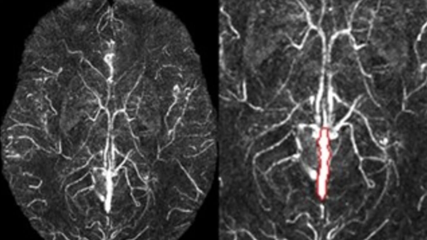
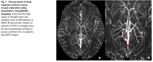
52 hemodialysis patients and 54 age-and gender-matched healthy controls were enrolled. QSM reconstructed from original phase data of 3.0 T susceptibility-weighted imaging was used to measure the susceptibility of straight sinus. The susceptibility was used to calculate the deep regional cerebral SvO2 and compare with healthy individuals. Correlation analysis was performed to investigate the correlation between deep regional cerebral SvO2, clinical risk factors and neuropsychiatric testing.
Results
The deep regional cerebral SvO2 of hemodialysis patients (72.5 ± 3.7%) was significantly lower than healthy controls (76.0 ± 2.1%) (P < 0.001). There was no significant difference in the measured volume of interests of straight sinus between hemodialysis patients (250.92 ± 46.65) and healthy controls (249.68 ± 49.68) (P = 0.859). There were no significant correlations between the measured susceptibility and volume of interests in hemodialysis patients (P = 0.204) and healthy controls (P = 0.562), respectively. Hematocrit (r = 0.480, P < 0.001, FDR corrected), hemoglobin (r = 0.440, P < 0.001, FDR corrected), red blood cell (r = 0.446, P = 0.003, FDR corrected), dialysis duration (r = 0.505, P = 0.002, FDR corrected) and parathyroid hormone (r = −0.451, P = 0.007, FDR corrected) were risk factors for decreased deep regional cerebral SvO2 in patients. The Mini-Mental State Examination (MMSE) scores of hemodialysis patients were significantly lower than healthy controls (P < 0.001). However, the deep regional cerebral SvO2 did not correlate with MMSE scores (P = 0.630).
Conclusions
In summary, the decreased deep regional cerebral SvO2 occurred in hemodialysis patients and dialysis duration, parathyroid hormone, hematocrit, hemoglobin and red blood cell may be clinical risk factors.

