Quantitative Susceptibility Mapping for Characterization of Intraplaque Hemorrhage and Calcification in Carotid Atherosclerotic Disease
By: Karen Holzberger, President & CEO of SpinTech MRI
Author(s): Chaoyue Wang, MSc,1,2,3 Yue Zhang, MS,1 Jingwen Du, MS,1 István N. Huszár, MD,2,3 Saifeng Liu, PhD,4 Yongsheng Chen, PhD,4,5 Sagar Buch, PhD,6 Fang Wu, MD,1 Yuehong Liu, MS,1 Mark Jenkinson, PhD,2,3 Charlie Chia-Tsong Hsu, PhD,7 Zhaoyang Fan, PhD,8 E. Mark Haacke, PhD,4,5 and Qi Yang, MD, PhD1,8*
Journal: Journal of Magnetic Resonance Imaging
Published: 2020
Read Full Paper: https://onlinelibrary.wiley.com/doi/abs/10.1002/jmri.27064
Abstract

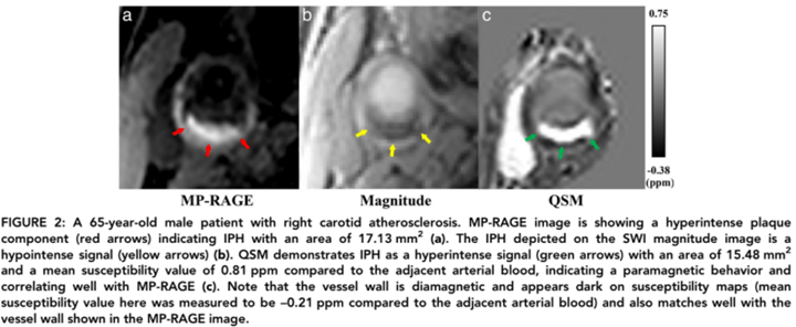
Carotid artery intraplaque hemorrhage (IPH), an unstable component of atherosclerosis, is associated with an increased risk of stroke. This prospective study aims to investigate the use of quantitative susceptibility mapping (QSM) as a tool for the evaluation of IPH and calcification in vivo. The study population included ten healthy volunteers and 15 patients.
Field Strength/Sequence
3.0T Susceptibility‐weighted imaging (SWI), magnetization‐prepared rapid acquisition with gradient echo (MP‐RAGE), T1‐weighted sampling perfection with application of optimized contrasts using different flip angle evolution (T1‐SPACE), T2‐weighted turbo spin‐echo (T2WI), and time‐of‐flight (TOF) sequences.
Assessment
The vessel wall area of the carotid artery was measured with QSM and compared with T1‐SPACE on healthy volunteers. Four radiologists, blinded to clinical history and patient identity, determined the presence and area of IPH on MP‐RAGE and QSM, as well as the area of calcification on T1‐SPACE and QSM.
Statistical Tests
Bland–Altman analysis, Pearson correlation coefficients, linear regression analyses were performed to evaluate the concordance of area measurements. Cohen’s kappa (κ) was analyzed to determine the agreement between IPH detections. The paired t‐test was used to compare the group differences.
Results
In 423 matched slices, 20.1% (85/423) and 19.6% (83/423) were detected to have IPH on MP‐RAGE and QSM, respectively. IPH detection by QSM and MP‐RAGE showed good agreement (κ = 0.822, P < 0.001) between the two methods. There was no significant difference in IPH area measurements between QSM and MP‐RAGE (7.28 mm2 ± 6.41 vs. 7.16 mm2 ± 5.99, P = 0.575). There was no significant difference in calcification area measurement between QSM and T1‐SPACE (3.51 mm2 ± 1.78 vs. 3.41 mm2 ± 2.02, P = 0.783).
Conclusion
QSM is a novel imaging tool for the identification of IPH in patients with carotid atherosclerosis and enables differentiation of IPH and calcification.

