Quantifying iron content in magnetic resonance imaging
By: Karen Holzberger, President & CEO of SpinTech MRI
Author(s): Kiarash Ghassaban a , Saifeng Liu b , Caihong Jiang c , E. Mark Haacke a,b,d,*
Journal: NeuroImage
Published: 2019
Read Full Paper: https://www.sciencedirect.com/science/article/abs/pii/S1053811918303574
Abstract

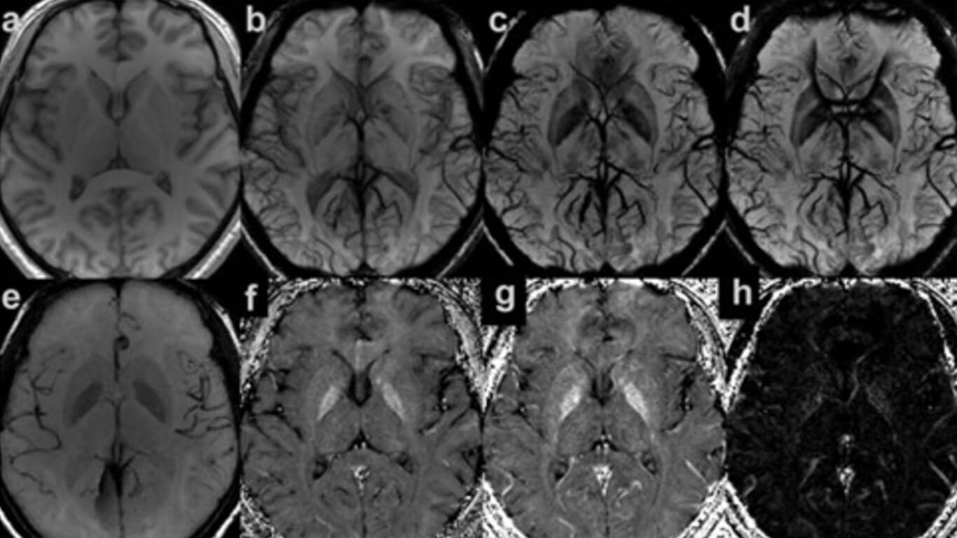
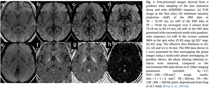
Measuring iron content has practical clinical indications in the study of diseases such as Parkinson’s disease, Huntington’s disease, ferritinopathies and multiple sclerosis as well as in the quantification of iron content in microbleeds and oxygen saturation in veins. In this work, we review the basic concepts behind imaging iron using T2, T2*, T2′, phase and quantitative susceptibility mapping in the human brain, liver and heart, followed by the applications of in vivo iron quantification in neurodegenerative diseases, iron tagged cells and ultra-small superparamagnetic iron oxide (USPIO) nanoparticles.

