Intracranial iron distribution and quantification in aceruloplasminemia: A case study
By: Karen Holzberger, President & CEO of SpinTech MRI
Author(s): Liche Zhoua,1 , Yan Chenb,1 , Yan Lic , Sara Gharabaghie , Yongsheng Chend , Sean K. Sethie,f , Yiwen Wua,⁎ , E.M. Haackea,e,f,⁎⁎
Journal: Magnetic Resonance Imaging
Published: 2020
Read Full Paper: https://www.sciencedirect.com/science/article/abs/pii/S0730725X19308094?via%3Dihub
Abstract

Aceruloplasminemia (ACP) is a rare autosomal recessive disorder characterized by intracranial and visceral iron overload. With R2*-based imaging or quantitative susceptibility mapping (QSM), it is feasible to measure iron in the brain quantitatively, although to date this has not yet been done for patients with ACP. The aim of this study was to provide quantitative iron measurements for each affected brain region in an ACP patient with the potential to do so in all future ACP patients. This may shed light on the link between brain iron metabolism and the territories affected by ceruloplasmin function.
Method
We imaged a patient with ACP using a 3T magnetic resonance imaging scanner with a fifteen-channel head coil. We manually demarcated gray matter and white matter on the Strategically Acquired Gradient Echo (STAGE) images, and calculated values for susceptibility and R2* in these regions. Correlation analysis was performed between the R2* values and the susceptibility values.
Results
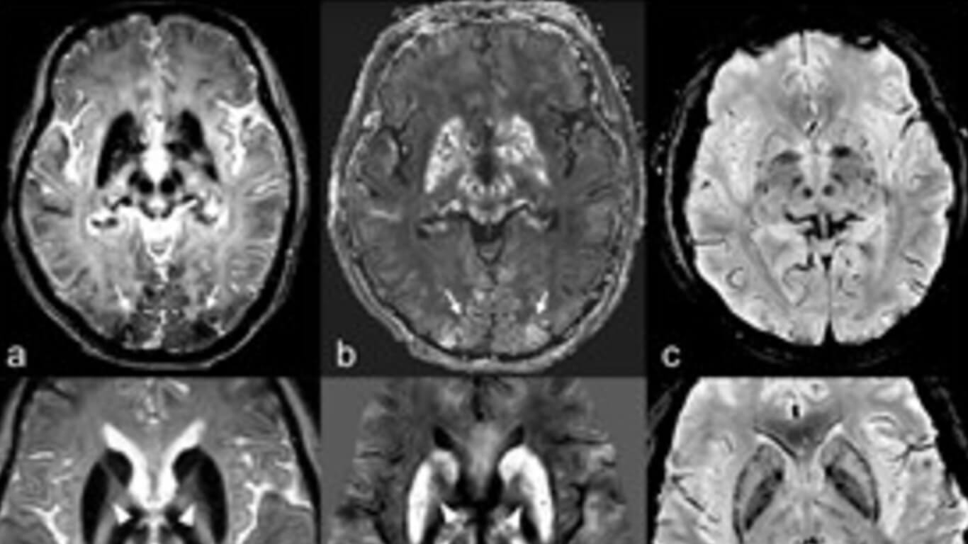
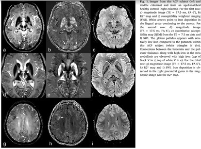
Besides the usual territories affected in ACP, we also discovered that the mammillary bodies and the lateral habenulae had significant increases in iron, and the hippocampus was severely affected both in terms of iron content and abnormal tissue signal. We also noted that the iron in the cortical gray matter appeared to be deposited in the inner layers. Moreover, several pathways between the superior colliculus and the pulvinar thalamus, between the caudate and putamen anteriorly and between the caudate and pulvinar thalamus posteriorly were also evident. Finally, R2* correlated strongly with the QSM data (R2 = 0.67, t = 6.78, p < 0.001).
Conclusions
QSM and R2* have proven to be sensitive and quantitative means by which to measure iron content in the brain. Our findings included several newly noted affected brain regions of iron overload and provided some new aspects of iron metabolism in ACP that may be further applicable to other pathologic conditions. Furthermore, our study may pave the way for assessing efficacy of iron chelation therapy in these patients and for other common iron related neurodegenerative disorders.

