Quantitative susceptibility-weighted imaging may be an accurate method for determining stroke hypoperfusion and hypoxia of penumbra
By: Karen Holzberger, President & CEO of SpinTech MRI
Author(s): Xiudi Lu1 & Linglei Meng2 & Yongmin Zhou3 & Shaoshi Wang2 & Miller Fawaz4 & Meiyun Wang5 & E. Mark Haacke4 & Chao Chai6 & Meizhu Zheng7 & Jinxia Zhu8 & Yu Luo3 & Shuang Xia6
Journal: European Radiology
Published: 2021
Read Full Paper: https://link.springer.com/article/10.1007%2Fs00330-020-07485-2
Abstract

Objectives
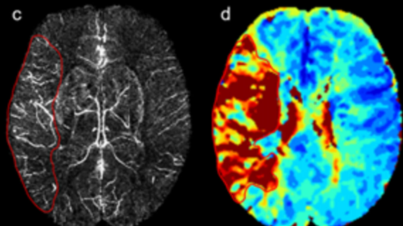
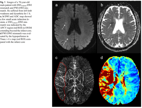
To quantitatively evaluate the volume of the ischemic penumbra using susceptibility-weighted imaging and mapping (SWIM) of asymmetrical prominent cortical veins (APCVs) in patients with acute ischemic stroke.
Methods
Eighty-five eligible patients with acute ischemic stroke on admission within 12 h from symptom onset were studied. The APCVs on SWIM were quantitatively (SWI-volume) and semi-quantitatively (SWI-Alberta Stroke Program Early CT Score, SWI-ASPECTS) evaluated to calculate mismatch. To assess the diagnostic efficacy of APCVs on SWIM, comparative analyses were performed between SWIvolume-DWI mismatch and SWIASPECTS-DWI mismatch, using PWI-DWI mismatch as a reference. Correlations were calculated between the mismatches, as well as between SWI-volume and time-to-maximum (Tmax) > 6 s volume. Additionally, each of these mismatches was correlated with the National Institute of Health Stroke Scale (NIHSS).
Results
The sensitivity, negative predictive value, and accuracy of SWIvolume-DWI mismatch were demonstrably higher than SWIASPECTS-DWI mismatch (100% vs. 53.7%, 100% vs. 9.5%, 97.7% vs. 54.5%, respectively). A significant positive correlation was found between SWIvolume-DWI and PWI-DWI mismatch (r = 0.691, p < 0.01), as well as between SWI-volume and Tmax > 6 s volume (r = 0.786, p < 0.001). A significant negative correlation was found between SWIvolume-DWI mismatch and NIHSS (r = − 0.360, p = 0.022), as well as between SWIASPECTS-DWI mismatch and NIHSS (r = − 0.499, p = 0.001).
Conclusions
SWIvolume-DWI mismatch had higher diagnostic efficacy than SWIASPECTS-DWI mismatch in defining the ischemic penumbra and showed good consistency with PWI-DWI mismatch in acute ischemic stroke. Quantitation of APCVs using SWIM provided an accurate method for determining hypoperfusion and provided a reliable method to reflect the hypoxia of penumbra.

