Multi-Echo Quantitative Susceptibility Mapping for Strategically Acquired Gradient Echo (STAGE) Imaging
By: Karen Holzberger, President & CEO of SpinTech MRI
Author(s): Sara Gharabaghi1,2, Saifeng Liu3, Ying Wang3,4, Yongsheng Chen5, Sagar Buch4, Mojtaba Jokar2, Thomas Wischgoll1, Nasser H. Kashou6, Chunyan Zhang7, Bo Wu8, Jingliang Cheng7 and E. Mark Haacke2,3,4,5,9
Journal: Frontiers in Neuroscience
Published: 2020
Read Full Paper: https://www.frontiersin.org/articles/10.3389/fnins.2020.581474/full
Abstract

The purpose of this study was to develop a method to reconstruct quantitative susceptibility mapping (QSM) from multi-echo, multi-flip angle data collected using strategically acquired gradient echo (STAGE) imaging.
Methods
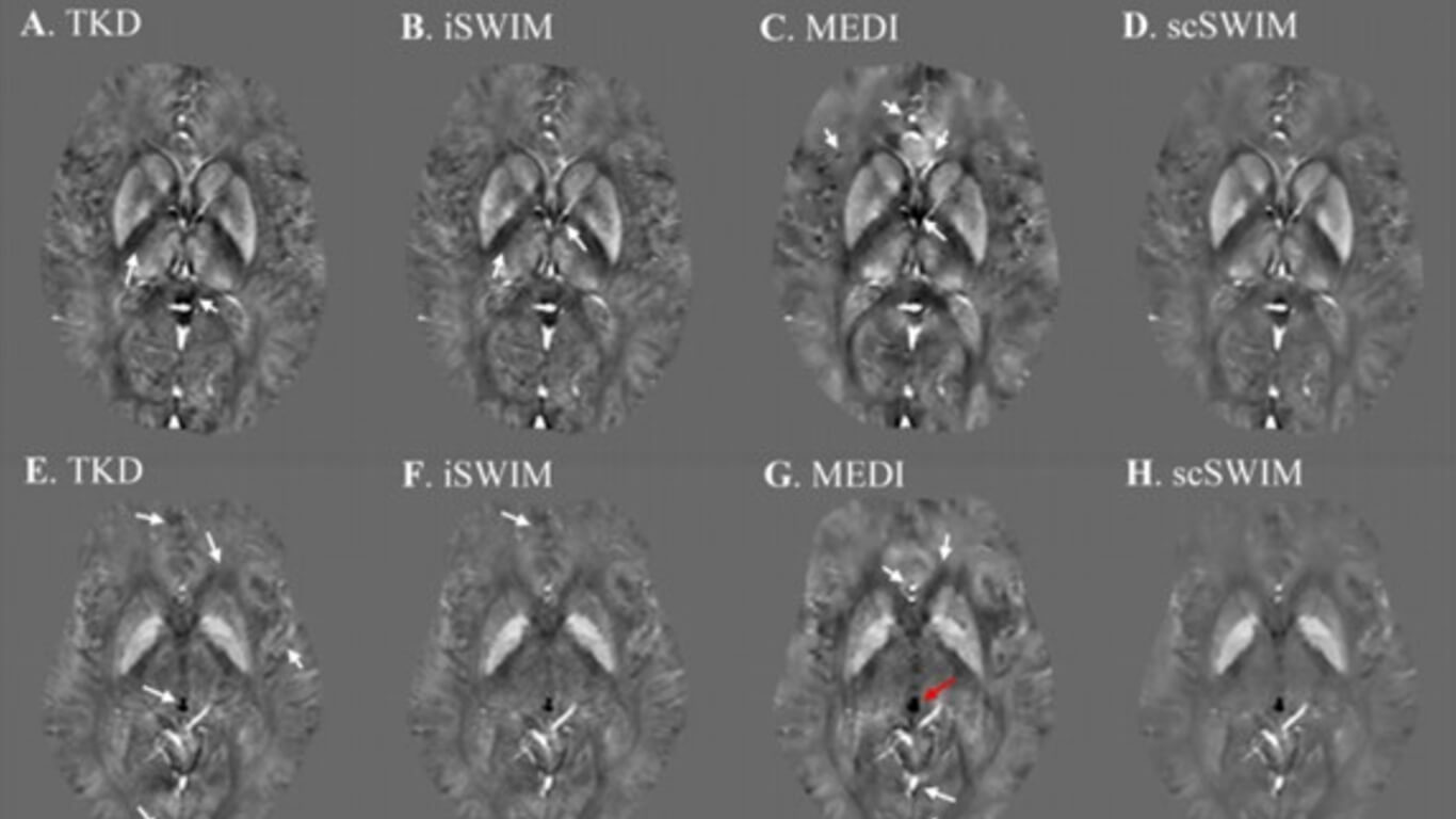
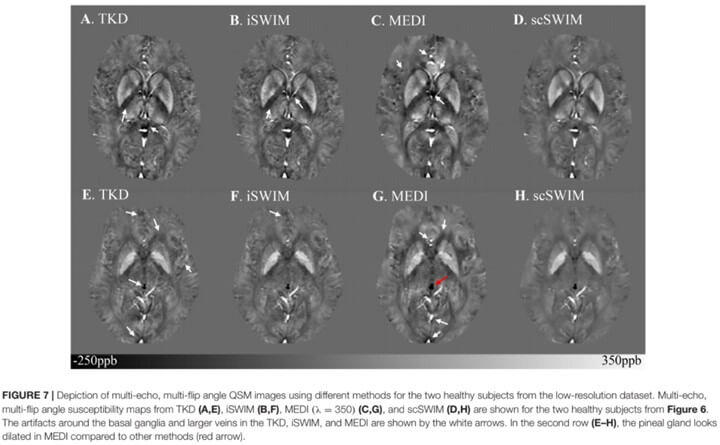
The proposed QSM reconstruction algorithm, referred to as “structurally constrained Susceptibility Weighted Imaging and Mapping” scSWIM, performs an ℓ1 and ℓ2 regularization-based reconstruction in a single step. The unique contrast of the T1 weighted enhanced (T1WE) image derived from STAGE imaging was used to extract reliable geometry constraints to protect the basal ganglia from over-smoothing. The multi-echo multi-flip angle data were used for improving the contrast-to-noise ratio in QSM through a weighted averaging scheme.
The measured susceptibility values from scSWIM for both simulated and in vivo data were compared to the: original susceptibility model (for simulated data only), the multi orientation COSMOS (for in vivo data only), truncated k-space division (TKD), iterative susceptibility weighted imaging and mapping (iSWIM), and morphology enabled dipole inversion (MEDI) algorithms.
Goodness of fit was quantified by measuring the root mean squared error (RMSE) and structural similarity index (SSIM). Additionally, scSWIM was assessed in ten healthy subjects.
Results
The unique contrast and tissue boundaries from T1WE and iSWIM enable the accurate definition of edges of high susceptibility regions. For the simulated brain model without the addition of microbleeds and calcium, the RMSE was best at 5.21ppb for scSWIM and 8.74ppb for MEDI thanks to the reduced streaking artifacts. However, by adding the microbleeds and calcium, MEDI’s performance dropped to 47.53ppb while scSWIM performance remained the same.
The SSIM was highest for scSWIM (0.90) and then MEDI (0.80). The deviation from the expected susceptibility in deep gray matter structures for simulated data relative to the model (and for the in vivo data relative to COSMOS) as measured by the slope was lowest for scSWIM + 1%(−1%); MEDI + 2%(−11%) and then iSWIM −5%(−10%). Finally, scSWIM measurements in the basal ganglia of healthy subjects were in agreement with literature.
Conclusion
This study shows that using a data fidelity term and structural constraints results in reduced noise and streaking artifacts while preserving structural details. Furthermore, the use of STAGE imaging with multi-echo and multi-flip data helps to improve the signal-to-noise ratio in QSM data and yields less artifacts.

