Assessment of Brain Venous Structure in Military Traumatic Brain Injury Patients using Susceptibility Weighted Imaging and Quantitative Susceptibility Mapping
By: Karen Holzberger, President & CEO of SpinTech MRI
Author(s): Wei Liu,1,2 Ping-Hong Yeh,1,2 Dominic E. Nathan,1,2 Chihwa Song,1 Helena Wu,1,2 Grant H. Bonavia,1 John Ollinger,1 and Gerard Riedy1
Journal: JOURNAL OF NEUROTRAUMA
Published: 2019
Read Full Paper: https://www.liebertpub.com/doi/10.1089/neu.2018.5970?url_ver=Z39.88-2003&rfr_id=ori%3Arid%3Acrossref.org&rfr_dat=cr_pub++0pubmed&

Abstract
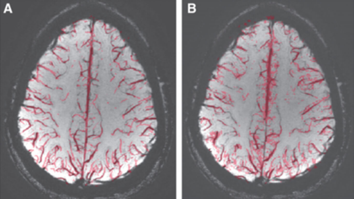
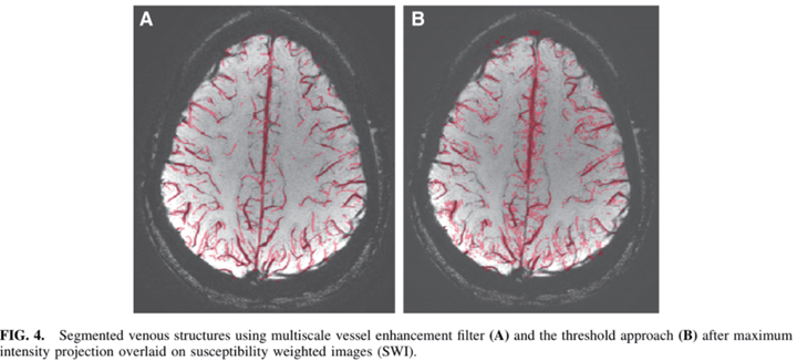
Brain venous volume above the lateral ventricle in military patients with traumatic brain injury (TBI) was assessed using two segmentation approaches on susceptibility weighted images (SWI) and quantitative susceptibility maps (QSM).
Method
This retrospective study included a total of 147 subjects: 14 patients with severe TBI; 38 patients with moderate TBI, 58 patients with mild TBI (28 with blast-related injuries and 30 with non-blast–related injuries), and 37 control subjects without history of TBI. Using the multiscale vessel enhancement filter on SWI images, patients with severe TBI demonstrated significantly higher segmented venous volumes compared with controls. Using a threshold approach on QSM images, TBI patients with different severities all demonstrated increased segmented volumes compared with control subjects: in the whole brain (severe, p = 0.001; moderate, p = 0.008; mild, p = 0.042, compared with controls), in the left hemisphere (severe, p = 0.01; moderate, p = 0.038, compared with controls), in the right hemisphere (severe, p = 0.001; moderate, p = 0.013; mild, p = 0.027, compared with controls).
Conclusions
While segmented volumes on SWI appear to overlay directly on the visualized venous structures, the QSM-derived segments also encompass some perivascular and deep white matter areas. This might represent the damage in the perivascular regions associated with iron deposition or astroglial scarring.

