Detection of hemorrhagic and axonal pathology in mild traumatic brain injury using advanced MRI: Implications for neurorehabilitation
By: Karen Holzberger, President & CEO of SpinTech MRI
Author(s): Randall R. Bensona,∗, Ramtilak Gattub, Bradley Sewickc, Zhifeng Koub, Nisrine Zakariahd, John M. Cavanaughd and E. Mark Haackeb
Journal: NeuroRehabilitation
Published: 2012
Read Full Paper: https://content.iospress.com/articles/neurorehabilitation/nre00795
Abstract

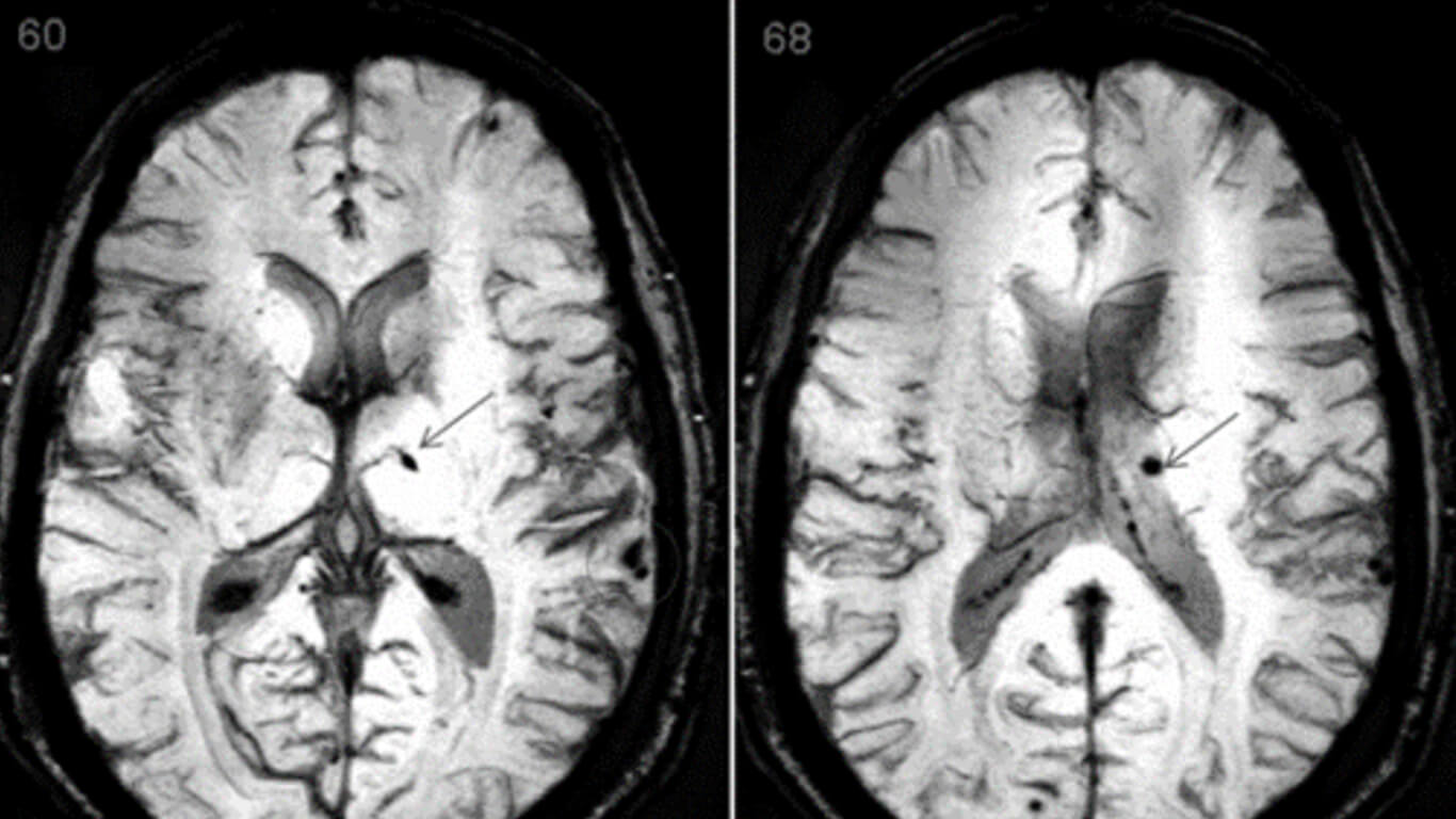
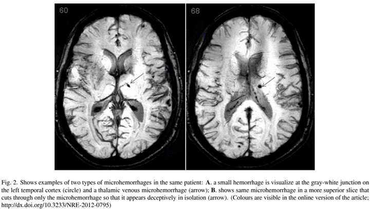
There is a need to more accurately diagnose milder traumatic brain injuries with increasing awareness of the high prevalence in both military and civilian populations. Magnetic resonance imaging methods may be capable of detecting a number of the pathoanatomical and pathophysiological consequences of focal and diffuse traumatic brain injury. Susceptibility-weighted imaging (SWI) detects heme iron and reveals even small venous microhemorrhages occurring in diffuse vascular injury. Diffusion tensor imaging (DTI) reveals axonal injury by detecting alterations in water flow in and around injured axons. The overarching hypothesis of this paper is that newer, advanced MR imaging generates sensitive biomarkers of regional brain injury which allows for correlation with clinical signs and symptoms.
Method
Studies involving subjects with a history of traumatic brain injury as well as healthy, non-trauma controls were used. Analysis involved comparison of TBI patients’ imaging results with healthy controls as well as correlation of imaging findings with clinical measures of injury severity. An additional animal study of Sprague-Dawley albino rats compared imaging results with histopathological findings after the animals were sacrificed and stained for b-APP.
Results
SWI revealed small foci of hemosiderin for some patients while aggregate lesion volume on SWI correlated with clinical injury severity indices. Similarly, DTI showed striking group differences for fractional anisotropy over the white matter globally, while tract and voxel-based regional results colocalized with SWI and FLAIR lesions in some cases and correlated with clinical deficits. For the rats, correlations were seen between imaging findings and staining of axonal injury.
Discussion
Animal data gave important tissue correlations with imaging results. SWI and DTI are commercially available sequences that can improve the diagnostic and prognostic ability of the trauma clinician. These biomarkers of regional brain injury which are present in imaging shortly after acute injury and persist indefinitely can inform clinicians and researchers about not only injury severity but also which neurobehavioral systems were injured. Analogous to stroke rehabilitation, having an understanding of the distribution of brain injury should ultimately allow for development of more effective rehabilitation strategies and more efficient clinical interventional trials.

