Vascular, flow and perfusion abnormalities in Parkinson’s disease
By: Karen Holzberger, President & CEO of SpinTech MRI
Author(s): Chunyan Zhanga,1 , Bo Wub,1 , Xiao Wanga , Chen Chena , Ruichen Zhaoa , Hong Luc , Hongcan Zhuc , Bing Xuec , Hong Liangc , Sean K. Sethid , E. Mark Haacked , Jinxia Zhue , Yuanyuan Penga , Jingliang Chenga,∗
Journal: Parkinsonism and Related Disorders
Published: 2020
Read Full Paper: https://www.prd-journal.com/article/S1353-8020(20)30048-1/fulltext
Abstract

Reduced flow into the brain or decreased jugular venous outflow from the brain may serve as a potential biomarker for Parkinson’s disease (PD). Our goal was to compare the presence of vascular abnormalities, flow, and increases in midbrain iron content (a hallmark of the disease) in patients with PD.
Methods
A total of 85 PD patients and 85 healthy controls (HCs) were imaged at 3T. We assessed vascular abnormalities using magnetic resonance (MR) venography, average cerebral blood flow using 2D flow quantification, and substantia nigra iron content using susceptibility mapping.
Results
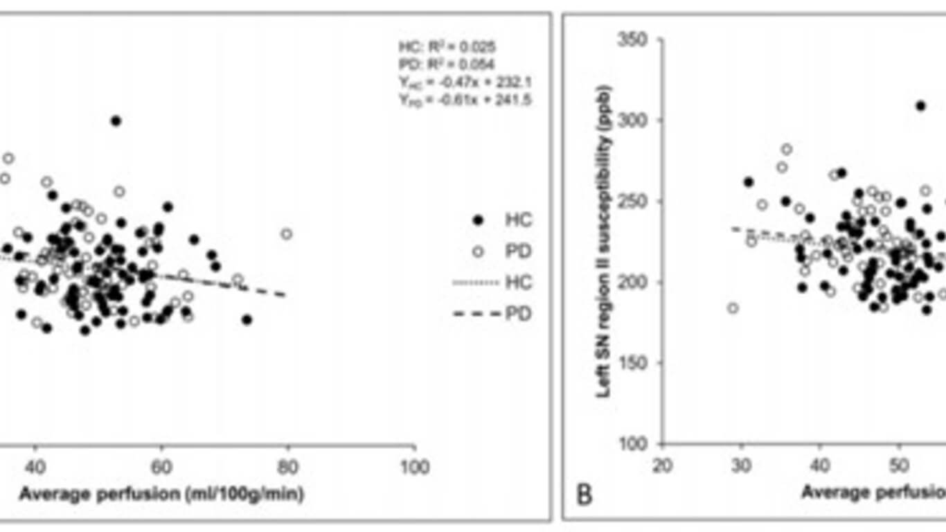
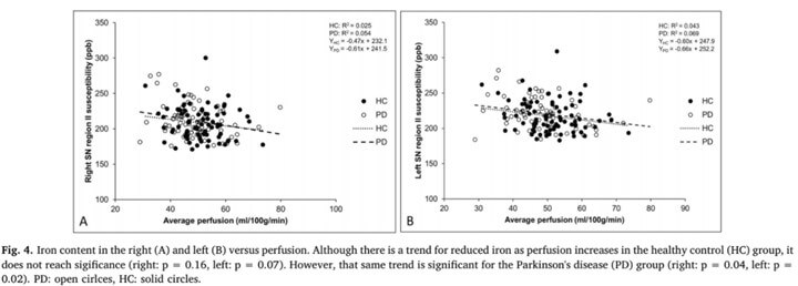
Fifty-two percent (52%) of the PD subjects showed venous structural and functional abnormalities in the two most severe categories, while ony 14% of the HC group showed these abnormalities. Total arterial flow (tA) was significantly lower for the PD group (10.9 ± 1.8 ml/s) compared to the HCs (11.6 ± 2.1 ml/s) (t = 2.28, p = 0.02). Of the PD patients (HCs), 42% (14%) had little flow on the left side. PD patients had higher heart rates and lower perfusion and the lower perfusion correlated with increased iron content in the substantia nigra.
Conclusion
Some PD patients showed abnormal left internal jugular veins, lower tA, higher heart rates, and lower perfusion relative to HCs. These indicators could serve as early biomarkers for PD and create new avenues for future studies regarding the etiology of PD.

