Quantitative measurements of brain iron deposition in cirrhotic patients using susceptibility mapping
By: Karen Holzberger, President & CEO of SpinTech MRI
Author(s): Shuang Xia1,2,*, Gang Zheng1,*, Wen Shen2 , Saifeng Liu4 , Long Jiang Zhang1 , E Mark Haacke5 and Guang Ming Lu1
Journal: Acta Radiologica
Published: 2015
Read Full Paper: https://journals.sagepub.com/doi/10.1177/0284185114525374?url_ver=Z39.88-2003&rfr_id=ori%3Arid%3Acrossref.org&rfr_dat=cr_pub++0pubmed&
Abstract

Susceptibility-weighted imaging (SWI) has been used to detect micro-bleeds and iron deposits in the brain. However, no reports have been published on the application of SWI in studying iron changes in the brain of cirrhotic patients.
The purpose of this study was to compare the susceptibility of different brain structures in cirrhotic patients with that in healthy controls and to evaluate susceptibility as a potential biomarker and correlate the measured susceptibility and cadaveric brain iron concentration for a variety of brain structures.
Method
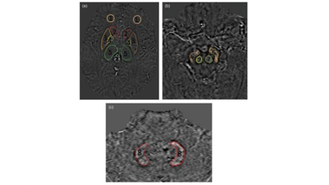
Forty-three cirrhotic patients (27 men, 16 women; mean age, 50 ± 9 years) and 34 age- and sex-matched healthy controls (22 men, 12 women; mean age, 47 ± 7 years) were included in this retrospective study. Susceptibility was measured in the frontal white matter, basal ganglia, midbrain, and dentate nucleus and compared with results gathered from two postmortem brain studies. Correlation between susceptibility and clinical biomarkers and neuropsychiatric tests scores was calculated.
Results
In cirrhotic patients, the susceptibility of left frontal white matter, bilateral caudate head, and right substantia nigra was higher than that in healthy controls (P < 0.05). There was a positive correlation between susceptibility and iron concentration from one postmortem brain study (r = 0.835, P = 0.01) in eight deep grey matter structures and another in five brain structures (r = 0.900, P = 0.03). The susceptibility of right caudate head (r = 0.402) and left caudate head (r = 0.408) correlated with neuropsychological test scores (both P < 0.05).
Conclusions
Abnormal iron deposits occur in cirrhotic patients and abnormal susceptibility of some brain regions appears to reflect neurocognitive changes.

