Susceptibility Weighted Imaging and Quantitative Susceptibility Mapping of the Cerebral Vasculature Using Ferumoxytol
By: Karen Holzberger, President & CEO of SpinTech MRI
Author(s): Saifeng Liu, PhD,1 * Jean-Christophe Brisset, PhD,2 Jiani Hu, PhD,3 E. Mark Haacke, PhD,1,3 and Yulin Ge, MD2
Journal: Journal of Magnetic Resonance Imaging
Published: 2017
Read Full Paper: https://onlinelibrary.wiley.com/doi/abs/10.1002/jmri.25809
Abstract

The purpose of this study was to demonstrate the potential of imaging cerebral arteries and veins with ferumoxytol using susceptibility weighted imaging (SWI) and quantitative susceptibility mapping (QSM).
Method
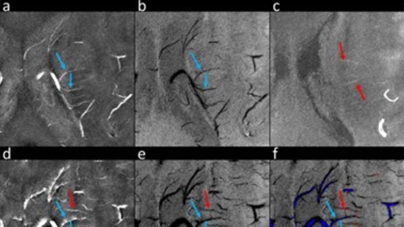
The relationships between ferumoxytol concentration and the apparent susceptibility at 1.5T, 3T, and 7T were determined using phantom data; the ability of visualizing subvoxel vessels was evaluated using simulations; and the feasibility of using ferumoxytol to enhance the visibility of small vessels was confirmed in three healthy volunteers at 7T(with doses 1 mg/kg to 4 mg/kg). The visualization of the lenticulostriate arteries and the medullary veins was assessed by two raters and the contrast‐to‐noise ratios (CNRs) of these vessels were measured.
Results
The relationship between ferumoxytol concentration and susceptibility was linear with a slope 13.3 ± 0.2 ppm·mg‐1·mL at 7T. Simulations showed that SWI data with an increased dose of ferumoxytol, higher echo time (TE), and higher imaging resolution improved the detection of smaller vessels. With 4 mg/kg ferumoxytol, voxel aspect ratio = 1:8, TE = 10 ms, the diameter of the smallest detectable artery was approximately 50μm. The rating score for arteries was improved from 1.5 ± 0.5 (precontrast) to 3.0 ± 0.0 (post‐4 mg/kg) in the in vivo data and the apparent susceptibilities of the arteries (0.65 ± 0.02 ppm at 4 mg/kg) agreed well with the expected susceptibility (0.71 ± 0.05 ppm).
Conclusions
The CNR for cerebral vessels with ferumoxytol can be enhanced using SWI, and the apparent susceptibilities of the arteries can be reliably quantified using QSM. This approach improves the imaging of the entire vascular system outside the capillaries and may be valuable for a variety of neurodegenerative diseases which involve the microvasculature

