Susceptibility mapping of the dural sinuses and other superficial veins in the brain
By: Karen Holzberger, President & CEO of SpinTech MRI
Author(s): Sagar Bucha,1 , Yongsheng Chenb , E. Mark Haackea,b,⁎
Journal: Magnetic Resonance Imaging
Published: 2019
Read Full Paper: https://www.sciencedirect.com/science/article/abs/pii/S0730725X18301607?via%3Dihub
Abstract

Quantitative susceptibility mapping (QSM) is a means to obtain direct measurements of local tissue susceptibility distribution. Usually the focus is on imaging tissues in the brain, and the region of the brain studied is dictated by an eroded skull stripped mask. Producing the pristine local phase behavior for regions at the edge of the brain has been difficult in the past.
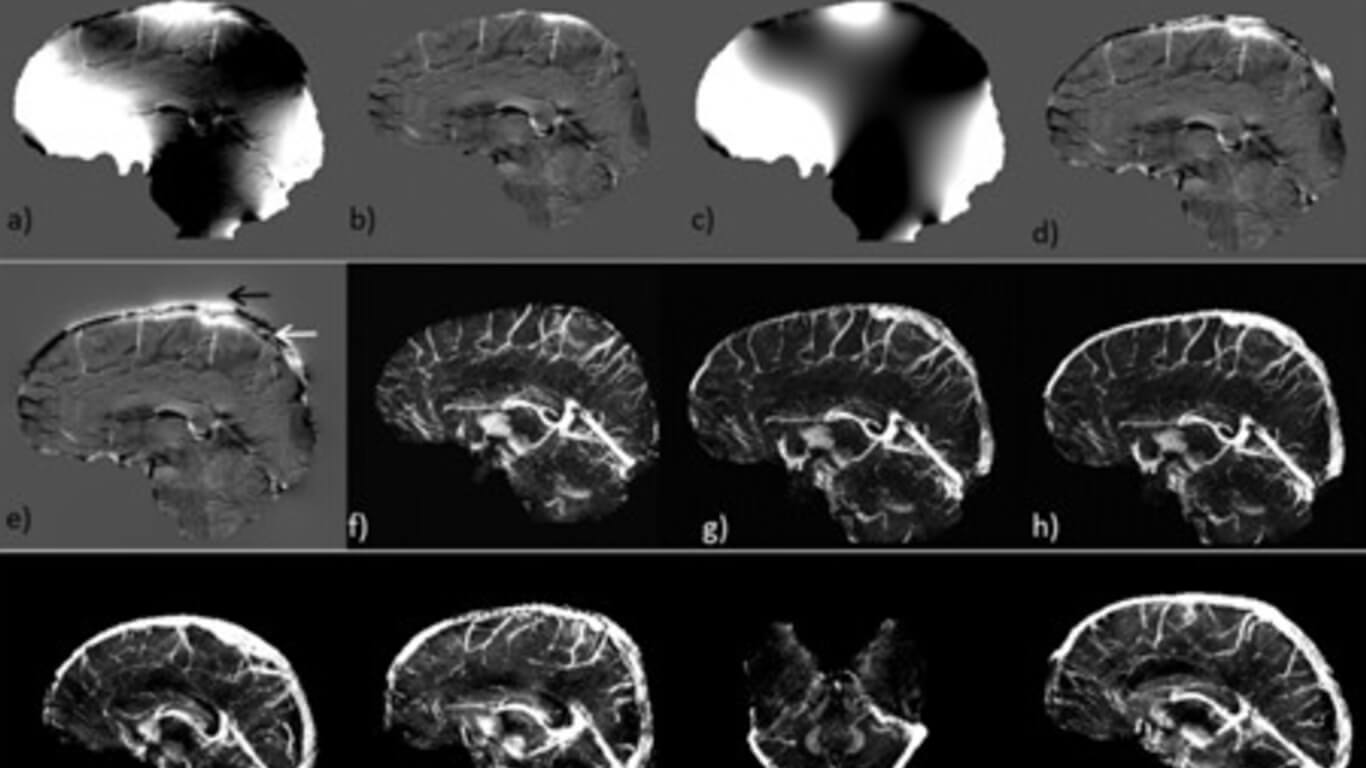
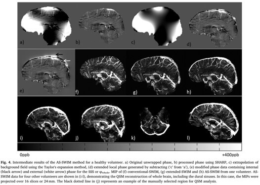
For structures such as the superior sagittal sinus (SSS) that run alongside the surface of the brain and under the skull bones, a considerable part of the external phase from the dipole effect is lost due to the short T2* of the bones. In this paper, we propose a method that seeks to reconstruct the susceptibility distribution inside the dural sinuses by ensuring that the entire geometry of the dural sinuses is preserved with the help of an MR angiogram and venogram (MRAV). Having a geometrical model of the vessels makes it possible to estimate the missing phase outside the brain as well, by using the forward phase model and, hence, allowing a complete phase map to be reconstructed.
Method
Fifteen healthy volunteers were scanned using a susceptibility weighted imaging (SWI) sequence with interleaved rephased-dephased echoes. QSM results were compared between the conventional techniques and the proposed method of phase preservation outside the brain and inside the dural sinuses.
Results
The mean and standard deviation inside the SSS for all volunteers was 435 ± 5 ppb (this is the inter-subject error). To validate the proposed approach, the mean susceptibility inside the straight sinus showed good agreement between conventional approach and the proposed method.
Conclusions
This method demonstrates the reconstruction of the SSS, whereas conventional methods are either unable to preserve this structure or unable to provide complete phase information. The results presented in this study indicate the potential of generating the susceptibility map for the whole brain, including the SSS (as well as potentially all the cortical veins).

