Quantifying Tissue Properties of the Optic Radiations Using Strategically Acquired Gradient Echo Imaging and Enhancing the Contrast Using Diamagnetic Susceptibility Weighted Imaging

Category: Papers

Author(s): P.K. Jella, Y. Chen, W. Tu, S. Makam, S. Beckius, E. Hamtaei, C.C.-T. Hsu, and E.M. Haacke
Journal: American Journal of Neuroradiology
Published: 2021
Read Full Paper: http://www.ajnr.org/content/early/2020/12/24/ajnr.A6897

Abstract
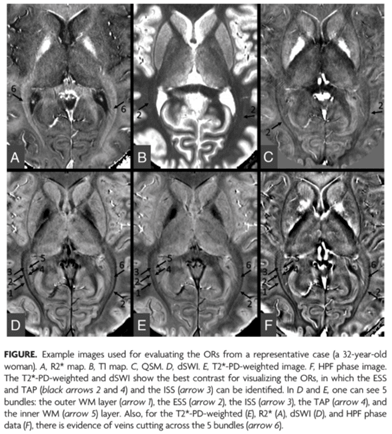
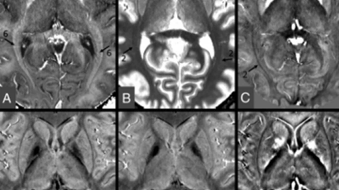
Visualization of the optic radiations is of clinical importance for diagnosing many diseases and depicting their anatomic structures for neurosurgical interventions. In this study, we quantify proton density, T1, T2*, and susceptibility of the optic radiation fiber bundles in a series of 10 healthy control participants using strategically acquired gradient echo imaging.

Furthermore, we introduce a novel means to enhance the contrast of the optic radiations using diamagnetic susceptibility weighted imaging.

New & Related

All Resources

Editorial: Quantitative Susceptibility Mapping in Neurodegeneration

Editorial: Quantitative Susceptibility Mapping in Neurodegeneration

The purpose of this study was to evaluate the effect of resolution on iron content using quantitative susceptibility mapping (QSM); to verify the consistency of QSM across field strengths and manufacturers in evaluating the iron content of deep gray matter (DGM) of the human brain using subjects from multiple sites; and to establish a susceptibility baseline as a function of age for each DGM structure using both a global and regional iron analysis.

read more
Iron Content in Deep Gray Matter as a Function of Age Using Quantitative Susceptibility Mapping: A Multicenter Study

Iron Content in Deep Gray Matter as a Function of Age Using Quantitative Susceptibility Mapping: A Multicenter Study

The purpose of this study was to evaluate the effect of resolution on iron content using quantitative susceptibility mapping (QSM); to verify the consistency of QSM across field strengths and manufacturers in evaluating the iron content of deep gray matter (DGM) of the human brain using subjects from multiple sites; and to establish a susceptibility baseline as a function of age for each DGM structure using both a global and regional iron analysis.

read more